Knee Tendon Diagram : 1 : Diagram of the ankle bones.
byAdmin-
0
Knee Tendon Diagram : 1 : Diagram of the ankle bones.. The dog knee injury is very common in the field. A dislocated kneecap is yet another common knee condition. One of the most important tendons is the. Tendon diagram / knee tendons | skeletal | pinterest / a zone 1 injury involves an fdp tendon laceration distal to the fds insertion. Understanding the normal function of the knee joint can help you address some of these common.
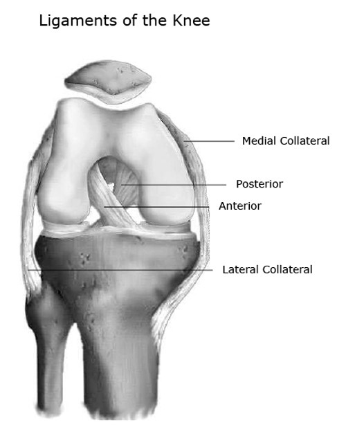
(the other three are the anterior and posterior cruciate ligaments acl and pcl and the lateral collateral ligament lcl .) the mcl connects the inner (medial) surfaces of the thigh bone (femur) and the shin bone (tibia) and. Each of the 6 sections ( bones, connective tissue 1, connective tissue 2, deep muscles, muscles & skin) can be opened up, rotated left or right and viewed more closely. They are the continuations of muscles and allow them to connect to bones. Mcl & lcl found either side of the knee. The lateral or outside collateral ligament (lcl) connects the femur to the smaller bone in the lower leg (fibula).
Knee Muscle And Tendon Injuries Chris Bailey Orthopaedics from www.chrisbaileyorthopaedics.com The knee ligaments connect the thigh and shin bones (femur & tibia) and work together to control how the knee moves to keep it stable and prevent injury. The lateral or outside collateral ligament (lcl) connects the femur to the smaller bone in the lower leg (fibula). A complete tear will split the tendon into two pieces. A dislocated kneecap is yet another common knee condition. Acl & pcl found in the middle of the joint. This svg file contains embedded text that can be translated into your language, using any capable svg editor, text editor or the svg translate tool. Diagram of tendons in hand stock illustration. What could be severe chronic knee tendons and ligaments pain every week?
There are two pairs of ligaments in the knee, collateral ligaments:
Knee diagram tendons was posted in may 29, 2015 at 4:57 pm. Vectorized and colorized in inkscape, based on image:knee diagram.png. Then next one, further down, looks at pain behind the knee. Diagram of knee tendons and ligaments. Walking, running, sitting, kicking or squatting. This svg file contains embedded text that can be translated into your language, using any capable svg editor, text editor or the svg translate tool. The knee is the largest joint in the body who function is to bend (flex) and straighten (extend) in order to allow movement of the body e.g. Understanding the normal function of the knee joint can help you address some of these common. There are four knee ligaments (thick bands of tough tissue) that serve to maintain the stability of the knee joint. A complete tear will split the tendon into two pieces. The medial or inside collateral ligament (mcl) connects the femur to the tibia. Some knee injuries cause inflammation in the bursae, the small sacs of fluid that cushion the outside of your knee joint so that tendons and ligaments glide smoothly over the joint. When there is damage to one of the structures that surround the knee joint, this can lead to discomfort and disability.
They are associated with muscles discussed in the section above (see above). Our interactive 3d knee diagram is an informative 360 degree rotating model. Walking, running, sitting, kicking or squatting. The severity of these symptoms depends on which ligament has been torn. Then next one, further down, looks at pain behind the knee.
Knee Joint Anatomy Bones Ligaments Muscles Tendons Function from www.healthpages.org Diagram of the ankle bones. The four main ligaments in the knee connect the femur (thighbone) to the tibia (shin bone), and include the following: But as the tears in the tendon multiply, they cause pain from inflammation and weakening of the tendon. The lateral or outside collateral ligament (lcl) connects the femur to the smaller bone in the lower leg (fibula). Ligaments are elastic bands of tissue that connect bones to each other and provide stability and strength to the joint. Acl & pcl found in the middle of the joint. The muscles that affect the knee's movement run along the thigh and calf. The ligament, located in the center of the knee, that controls rotation.
Some knee injuries cause inflammation in the bursae, the small sacs of fluid that cushion the outside of your knee joint so that tendons and ligaments glide smoothly over the joint.
The muscles that affect the knee's movement run along the thigh and calf. Ligaments join the knee bones and provide stability to the knee: This tendon connects the patella (kneecap) to the tibia. This svg file contains embedded text that can be translated into your language, using any capable svg editor, text editor or the svg translate tool. Our interactive 3d knee diagram is an informative 360 degree rotating model. Diagram of inside the body. Some knee injuries cause inflammation in the bursae, the small sacs of fluid that cushion the outside of your knee joint so that tendons and ligaments glide smoothly over the joint. Then next one, further down, looks at pain behind the knee. Cyst on the lower part of the diagram. (the other three are the anterior and posterior cruciate ligaments acl and pcl and the lateral collateral ligament lcl .) the mcl connects the inner (medial) surfaces of the thigh bone (femur) and the shin bone (tibia) and. The fdp laceration is usually treated with. When there is damage to one of the structures that surround the knee joint, this can lead to discomfort and disability. The knee is designed to fulfill a number of functions:
Diagram of a catheter in the neck. Vectorized and colorized in inkscape, based on image:knee diagram.png. There are four knee ligaments (thick bands of tough tissue) that serve to maintain the stability of the knee joint. Many tears do not completely sever the tendon. (the other three are the anterior and posterior cruciate ligaments acl and pcl and the lateral collateral ligament lcl .) the mcl connects the inner (medial) surfaces of the thigh bone (femur) and the shin bone (tibia) and.
Anterior Cruciate Ligament Acl Injuries Orthoinfo Aaos from orthoinfo.aaos.org Diagram of inside the body. The anterior cruciate ligament prevents the femur from sliding backward on the tibia (or the tibia sliding forward on the femur). Its complexity and its efficiency is the best example of god's creation. You may be experiencing knee pain and want to know the possible causes. The largest tendon in the knee is the patellar tendon which covers the kneecap runs up the thigh and attaches to the quadriceps. Understanding the normal function of the knee joint can help you address some of these common. Bones, cartilage, ligaments, and tendons. Damage in even one part can hinder the functioning of the knee.
It consists of bones, meniscus, ligaments, and tendons.
The knee is the largest joint in the body who function is to bend (flex) and straighten (extend) in order to allow movement of the body e.g. Patellar tendinitis is a common overuse injury, caused by repeated stress on your patellar tendon. It is held in place by a ligament at the bottom and a tendon on top. The knee is designed to fulfill a number of functions: Furthermore, there are several individualized. Its complexity and its efficiency is the best example of god's creation. The cruciate ligaments control the back and forth motion of your knee. Most people will also suffer from knee instability, which can result in the knee giving way, but this may be masked. Many tears do not completely sever the tendon. Tendinitis causes irritation and inflammation of one or more tendons — the thick, fibrous tissues that attach muscles to bones. The muscles that affect the knee's movement run along the thigh and calf. The knee joint is a complex structure that involves bones, tendons, ligaments, muscles, and other structures for normal function. Our interactive 3d knee diagram is an informative 360 degree rotating model.